What is the difference between spectral, multispectral, and hyperspectral
Spectral, multispectral, hyperspectral, can't tell the difference? 
Spectral analysis as an important means of natural science analysis, spectral technology is often used to detect the physical structure of objects, chemical composition and other indicators. Image spectrometry, on the other hand, combines spectral technology and imaging technology, combining spectral resolution capability and graphic resolution capability, resulting in faceted spectral analysis in the spatial dimension, which is now known as multispectral imaging and hyperspectral imaging technology.
What is the difference between spectral, multispectral and hyperspectral?
Spectrum
Spectrum is the monochromatic light separated by dispersion after the dispersive system (such as prisms, gratings), through the imaging system, projected on the detector to become the wavelength (or frequency) size of the sequential arrangement of the pattern, which is known as the optical spectrum. Ocean Optics spectrometer is based on this principle of design and manufacture.
Light waves according to different wavelengths, there are different names: wavelengths in the 380 and 780nm between the light waves known as visible light, shorter than 380nm called ultraviolet light; and longer than 780nm for the infrared light (infrared light is also divided into the near-infrared, mid-infrared, far-infrared, etc.).
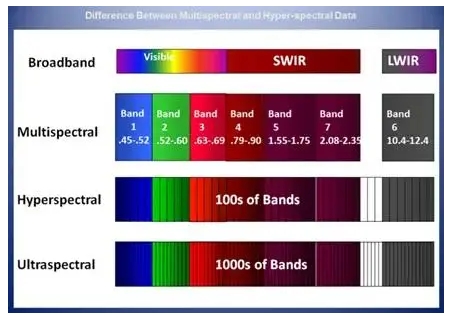
Multispectral technology refers to the simultaneous acquisition of multiple optical spectral bands (usually greater than or equal to 3), and in the visible light on the basis of infrared light and ultraviolet light to expand the direction of the spectral detection technology. The common realization method is through a variety of filters or beam splitters and a variety of combinations of photographic film, so that at the same time, respectively, to receive the same target in a range of different narrow spectral bands of light signals radiated or reflected, to get the target in several different spectral bands of the photo. The most common multispectral photos around are those taken by color cameras, as shown below, which contain information in three optical spectral bands, red (1), green (2) and blue (3), from the spectral point of view. If more bands are added to the camera or detector, such as bands (4) and (5), a multispectral photo with multiple bands can be obtained.
Multi-spectral technology combined with imaging hardware allows multi-spectral information to be presented in image form.
Of course, it is also possible to use only the detector to obtain the spectral information of a single spatial point. Pixelteq, a brand of Ocean Optics, with its unique chip filtering technology, can realize the acquisition of 8 channels of spectral information on a 9*9cm chip, which is especially suitable for applications with extremely high space and cost requirements.
Hypespectral is a fine technology that can capture and analyze the spectra point by point in a spatial area, due to the unique spectral “features” that can be detected in different spatial locations of a single object, and therefore can detect substances that cannot be distinguished visually.
Hyperspectral example: images consist of narrower bands (10-20 nm). Hyperspectral images may have hundreds or thousands of bands. After an object interacts with light from a light source and is received by a non-imaging spectral analysis device (e.g., a spectrometer), the device can accurately react to the intensity differences in the distribution of the received light signal over the spectral bands also known as spectral information. When using hyperspectral equipment, from the perspective of imaging characteristics, you can understand the spectral information of each position of the sample, from the perspective of spectral characteristics, you can understand the distribution of the signal position in a specific spectral band, that is, hyperspectral equipment can obtain richer detail information. For example: the human eye can only receive three spectral bands in the object's light energy signal: red, green and blue. That is, we often referred to as the three primary colors, but in fact we can see the combination of these three colors produced by the orange, purple, lime green and so on the more subtle colors. However, we are not able to distinguish the difference between pure yellow and a mixture of red and green, which is also known as “isochromatic”. But hyperspectral imaging can easily distinguish the difference.
Above, the two yellows, one a “solid color” and the other a mixture of red and green, may be visually indistinguishable, but due to their spectral differences, they can be distinguished using spectroscopic equipment. In our experiments, the data obtained with a spectrometer represent the average of the light emitted by all the molecules interacting with the incident light source over the entire detected range, whereas with a multispectral device it is possible to obtain information about the samples in a few specific bands at various points within the detected range. As a result, neither of these devices can provide very fine sample information in a single region.
A hyperspectral imager (HSI) can be analogized to hundreds or thousands of single-point spectrometers lined up closely together and focusing on an area at the same time, with each spectrometer working independently and acquiring spectral information about its own location. The data output from HSI is an image, or video stream, in which each pixel has its own spectrum, and each spectrum contains hundreds of spectral bands. This “full-spectrum” capability of hyperspectral imaging allows one to see the spectral signals at every distinguishable spatial location in a scene, i.e., more dimensional information is obtained. Therefore, hyperspectral imaging can be used in a variety of applications, including artwork identification, crop health, coastline mapping, forestry, mineral exploration, urban and industrial infrastructure, product quality in production lines, environmental monitoring, and more.
Hyperspectral scanning methods and imaging results

The difference between hyperspectral and multispectral
Very often the reflectance characteristic spectrum of a material can be very complex with respect to wavelength, and other minute features may not be distinguishable using coarser multispectral imaging methods.

Substances that were indistinguishable from those identified using multispectral imaging (left) in the figure above were distinguished through the use of hyperspectral imaging (right). The reason for this is that because hyperspectral has more spectral bands, more complex fingerprint features can be accurately obtained with higher spectral resolution.
Typical Applications
Hyperspectral devices can detect specific paints or dyes in the infrared that are not visible to the human eye. Similarly, HSI systems in the 60 or 300 band can provide richer spectral information on the reflectance of a material than a multispectral system, allowing for more accurate material characterization. The image below shows the image and spectral information obtained from a piece of fresh animal tissue placed on a conveyor belt in a laboratory using a hyperspectral imager:

Spectrograms of different regions: (a) labeled regions of pure fat, marbling and pure lean portions on tissue samples; (b) Spectrograms labeled in different regions of the (a) diagram.
In addition we can provide intuitive software programs for imaging analysis, classification and visualization of different substances with unique spectral characteristics. Whether this data is obtained from the air, on the ground, or in the lab, you can see details on your computer screen that may not be distinguishable by eye.


How can we Help you?
Email*
Phone*
Name*
Country/Region*
Which Products are you interested in?*